

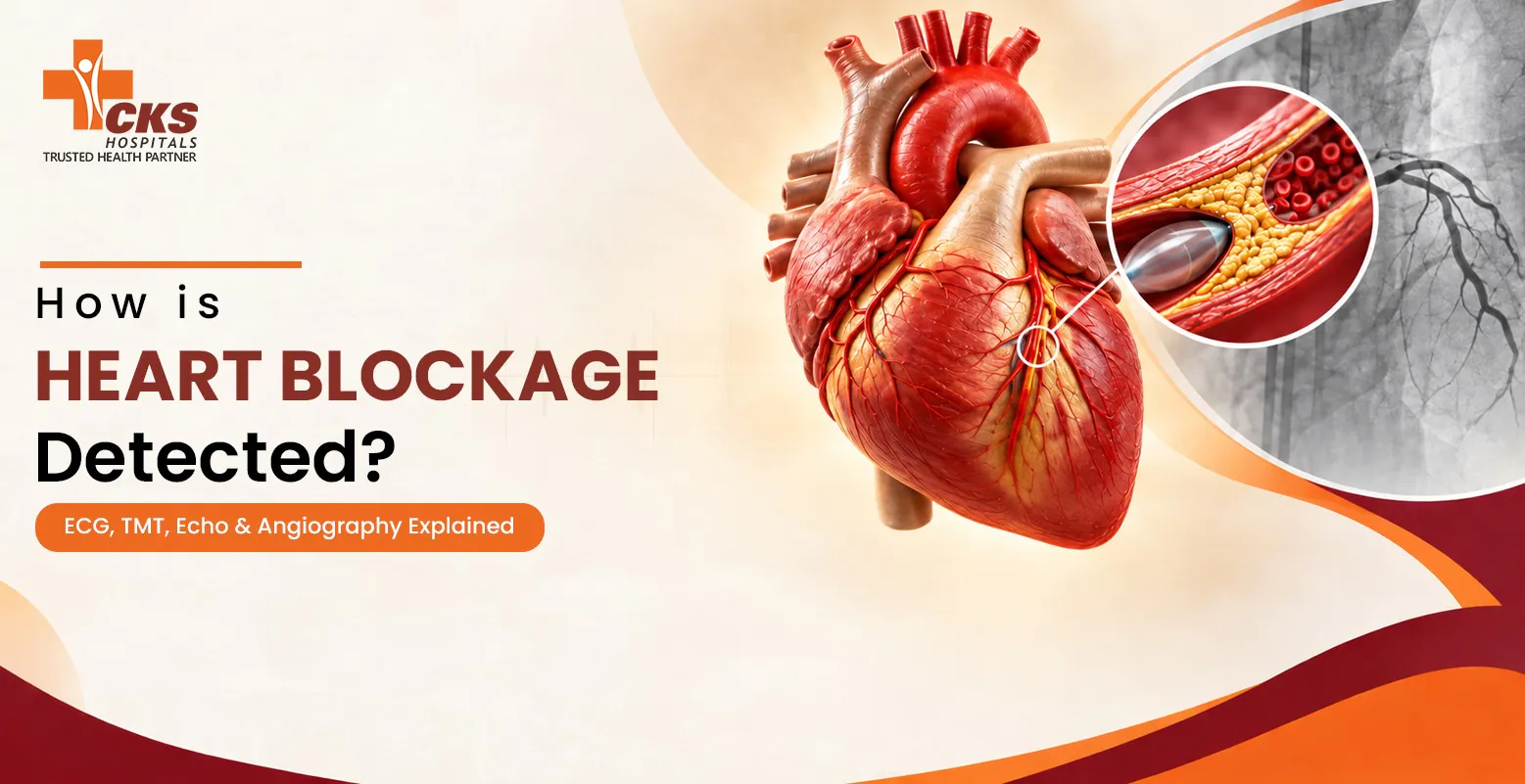
How Is Heart Blockage Detected? ECG, TMT, Echo & Angiography Explained
Do you sometimes feel pressure or tightness in your chest while walking or climbing stairs? Both can be symptoms of severe cardiac inflammation and heart blockage. To diagnose such cardiovascular conditions, your doctor may recommend some specific tests. One such group of tests is heart blockage detection, this test is used to find narrowed coronary arteries, plaque buildup and measure blood flow. It is a series of tests that includes an ECG, TMT, echocardiogram or coronary angiography.
Each of the above tests provides different insights about the functioning of your cardiovascular system and not every patient may need all of them. On that note, this guide explains how doctors use these tests, what heart blockage is, the difference between each test and when you should consult a cardiologist.
Medical Note: Heart blockage can have different causes and not all of them may develop noticeable symptoms. Diagnostic tests such as TMT, ECG, echocardiography and coronary angiography are used to assess different aspects of heart health. So, a cardiologist may order tests depending on the symptoms you feel and your medical history.
What Is Heart Blockage and Why Does It Need Timely Detection?
A heart blockage (also known as coronary artery disease) occurs when one or multiple coronary arteries narrow or become completely blocked because of the buildup of fatty deposits, cholesterol, plaque and calcium. The buildup of these substances affects the flow of oxygen-rich blood to the heart muscle. Because of that, a person who has a heart blockage may feel chest pain, and sometimes it leads to heart attacks and other serious cardiovascular complications.
It is also important to note that people sometimes confuse “Heart Blockage” with “Heart Block”. Both of them sound similar, but they are different conditions. As mentioned, heart blockage is the narrowing or complete blockage of coronary arteries because of the accumulation of some substances. On the other hand, a heart block is an electrical condition that causes a slow or irregular heartbeat.
Common Warning Symptoms
The symptoms of heart blockage may vary from person to person. The common warning signs of this condition are chest pain, pressure or tightness. Some people may also experience the following symptoms:
- Breathlessness
- Discomfort that spreads to the shoulder, arm, neck, jaw or back
- Dizziness
- Nausea
- Unusual tiredness
People who experience recurrent chest discomfort should consult a doctor right away for the most appropriate chest pain treatment.
Why Early Detection Matters?
Early detection of heart blockage through ECG, TMT, echo, and angiography is useful for identifying reduced blood flow and determining whether further testing is needed. A timely medical assessment also allows doctors to assess your overall cardiovascular system as well as related conditions such as high blood pressure or high cholesterol.
If symptoms suggest a possible coronary problem, coronary artery blockage diagnosis may include more than one type of test. The actual number of tests depends on your symptoms and medical history.
Step 1: ECG (Electrocardiogram), the First Screening Test
An electrocardiogram (ECG) test is usually done when a physician suspects heart problems based on symptoms experienced. This test provides the physician with a quick way of finding out whether heart disease is causing the symptoms and whether more tests are required to diagnose the condition.
What does an ECG Measure?
An electrocardiogram (ECG) is an instrument for recording electrical activity of the heart using small electrodes attached to the skin of the chest and arms of a patient. An ECG test measures heart rate and rhythm and detects some abnormalities in the electrical activity of the heart.
What Can and Cannot an ECG Tell You About a Blockage?
If you are wondering how is heart blockage detected through an ECG, it is important to note that this test’s results do not directly show the coronary arteries. ECG can sometimes show changes suggesting that the heart muscles are not receiving an adequate amount of blood or have been affected by a previous heart attack. However, an ECG by itself cannot specifically detect the precise location or severity of a coronary artery blockage.
When a Normal ECG Doesn’t Rule Out Blockage?
The ECG in the resting state may show a normal reading despite a coronary blockage. This happens because the severity of the blockage does not affect the heart’s electrical activity significantly at the time of rest. If further symptoms persist to suggest less blood supply to the heart muscles, then additional heart blockage tests may be ordered by your doctor for an accurate assessment.
Step 2: TMT (Treadmill Test), Testing the Heart Under Exertion
A TMT is useful for checking how your heart responds when it has to work under higher pressure than at rest. The test gradually increases the exercise intensity for a clearer analysis of heart function.
How does the TMT Procedure Work?
During a TMT, some electrodes are placed on your chest to continuously record your heart’s electrical activity. The TMT test begins with normal walking on the treadmill, after a few minutes, the speed and incline are gradually increased. Because the exercise intensity rises, your heart rate may also increase.
At this point, your heart rate, blood pressure and symptoms are monitored throughout the test. If you experience significant chest discomfort or breathlessness, the test can be stopped.
Why Exertion Reveals What a Resting ECG Misses?
While you exercise, the heart needs more oxygen-rich blood. If the coronary arteries are significantly narrowed, the rising demand for blood may cause symptoms or ECG changes. These symptoms are often not present when you are at rest. This whole process makes a TMT test for heart blockage useful for patients who are being evaluated for possible reduced blood flow to the heart.
Limitations of TMT
The core limitation of the TMT is that it does not show visuals of coronary arteries or precisely locate a blockage. The results of this test can sometimes be inconclusive or affected by other heart conditions. This means that an abnormal test result does not automatically confirm severe heart blockage. Depending on the overall assessment, your doctor may recommend additional heart blockage tests.
Step 3: Echocardiogram (Echo) – Visualising Heart Structure and Function
An echocardiogram test allows doctors to visually confirm how well the heart is working. This test can be used alongside other tests that are used to diagnose cardiovascular conditions for more accuracy.
How Echo Imaging Works?
An Echo test for the heart uses high-frequency sound waves to create heart images on a screen. It operates on the same basic principle as the sonar used by submarines to navigate. During the test, a special gel is applied over the skin of the chest area, and a handheld device called a transducer is gently moved over the same areas to generate a heart image.
Stress Echo as an Enhanced Option
A stress echocardiogram indicates how the heart works under intense load. This test for heart blockage detection is often performed by exercising on a treadmill or using medicines that increase the frequency of heartbeat. The doctor captures ultrasound images of the heart before and during the stress to record changes that occur in the cardiovascular system.
If some areas of the heart do not move as expected during the test, this could mean that they are not getting sufficient blood.
What Echo Reveals vs What It Can’t
An echocardiogram can indicate how well your heart is pumping blood and whether there are any concerning problems with the heart valves and movement of the heart muscles. These results are also useful when a doctor is assessing conditions that require heart failure treatment.
An echocardiogram cannot provide visuals of coronary arteries or a detailed picture of a blockage. To confirm the actual cause of the condition, doctors may recommend additional tests.
Coronary Angiography – The Gold Standard for Detecting Blockage
Coronary angiography allows the cardiologist to visualise the arteries that supply the heart with blood. The images show the points of any narrowing of the arteries and will help the doctor determine the effect that it may have on the flow of blood.
How the Catheter and Dye Procedure Works?
For angiography, the doctor inserts a thin and flexible catheter into the blood vessel through either the wrist or groin. The catheter will then be moved to the heart and a contrast dye will be injected into it. The X-ray pictures will be taken as the contrast dye moves through the coronary arteries, making the arteries more visible. This angiography test for heart blockage typically takes place in a catheterisation laboratory.
CT Angiography vs Invasive Angiography
With a Computed Tomography (CT) angiography, the procedure is relatively simple and non-invasive. A standard intravenous (IV) catheter is placed into a vein in your arm. Once the contrast dye is injected through this arm vein, the actual scanning process takes only a few seconds while you lie on a table that moves through a doughnut-shaped scanner.
On the other hand, invasive angiography is a catheter-based surgical procedure performed in a specialised hospital suite. For this test, a doctor makes a small puncture in the femoral artery in the groin or the radial artery in the wrist. A thin, flexible tube called a catheter is then threaded through the arterial system and guided by continuous X-ray imaging. Both of these tests provide useful insights for coronary artery blockage diagnosis. Using any of them depends on your symptoms, health history, risk factors and earlier test results.
Why Angiography Confirms and Locates Blockage Precisely?
An angiography is able to confirm and locate blockage precisely because it combines a direct injection of radiopaque contrast dye with real-time X-ray fluoroscopy. This combination is useful for creating a detailed visual roadmap of blood flow through the arteries.
The Diagnostic Pathway: From Symptom to Confirmed Blockage
How is heart blockage detected depends on the symptoms as well as the medical history of each patient. The diagnosis typically begins with a clinical assessment and an ECG. Some additional tests may also be recommended, such as TMT and an echocardiogram.
Once these results are collected, the cardiologist decides if further testing is necessary. If the results indicate problems in the coronary arteries, the doctor recommends a CT coronary angiography or an invasive angiography. This step-by-step approach makes the whole heart blockage detection process more accurate and it helps doctors to guide the right treatment for each patient.
ECG vs TMT vs Echo vs Angiography: A Quick Comparison
ECG, TMT, Echo and angiography each help doctors understand your heart from a different perspective. The comparison below explains what all heart blockage tests check, how they are performed, how long they take and what preparation you may need:
| Test | Purpose | Accuracy | Invasiveness | Cost in India | Duration | Preparation Needed |
| ECG | Checks heart rhythm and electrical activity | Useful for detecting certain heart changes, but does not directly show blockages | Non-invasive | Usually low | 5–10 minutes | Usually none |
| TMT | Checks the heart’s response to exercise | Useful in selected patients; results are interpreted with other findings | Non-invasive | Usually moderate | 15–30 minutes | Comfortable clothing; follow doctor’s instructions |
| Echo | Shows heart structure, movement and pumping function | Provides detailed information about heart function | Non-invasive | Usually moderate | 20–45 minutes | Usually none |
| Angiography | Shows the coronary arteries and areas of narrowing | Provides a detailed view of coronary artery narrowing | CT is non-invasive; invasive angiography uses a catheter | Varies by type and hospital | Usually 30–60 minutes for invasive testing | May require fasting and other instructions |
The right test depends on your symptoms, medical history and earlier findings. Heart blockage detection may involve one test or a combination of tests.
Which Heart Test Do You Actually Need?
The right heart blockage tests depend on your symptoms and your history. The following recommendations are given according to the specific circumstances of each patient:
For a Routine Health Checkup
First, the doctor performs an evaluation of the heart and other risk factors. An electrocardiogram test can be recommended as an initial assessment. Further testing may be needed based on the outcomes of the diagnostics.
For Chest Pain or Breathlessness on Exertion
Chest discomfort or breathlessness during normal activity is worth getting checked by a doctor. These symptoms of heart blockage may lead your doctor to order an ECG, TMT, Echo or other tests as per your health profile.
For Patients With Known Risk Factors
Risk factors include high blood pressure, high cholesterol levels, diabetes, as well as family history of heart disease. Depending on your risk profile, your doctor may recommend that you undergo a heart checkup.
When Should You See a Cardiologist for Heart Blockage?
You should consult a cardiologist if you experience sudden breathing issues, severe chest pain, fainting and a sudden increase in heartbeat. A prompt medical assessment can help you get the most appropriate treatment plan.
Red-Flag Symptoms That Need Urgent Evaluation
You should seek urgent medical attention if you experience any of the following symptoms:
- Severe or sudden chest pain or discomfort
- Shortness of breath
- Chest discomfort with cold sweats
- Fainting or near fainting
- Chest pain or discomfort extending to the arm, jaw, back or shoulder
- Symptoms that persist or become rapidly worse
What to Expect During Your First Cardiology Consultation?
Your first cardiology consultation begins with telling your symptoms and medical history to the doctor. During the assessment, the cardiologist may also ask you about similar health conditions in your family, whether you are taking any medicines and may perform a physical examination.
Based on the initial findings, they may recommend a specific test or multiple tests. You can expect a similar process for a first consultation with the best cardiologist for heart blockage in Jaipur.
Why Choose Dr. Prakash Chandwani for Heart Blockage Detection and Treatment?
Dr. Prakash Chandwani is regarded as the best cardiologist in Jaipur and has over 25 years of experience in cardiology and coronary intervention.
The following are some of the major points regarding his clinical expertise and experience:
25+ Years of Experience in Interventional Cardiology
Dr. Prakash Chandwani is an Interventional Cardiologist at CKS Hospital, Jaipur. He has 25 + years of experience in cardiology. His qualifications include MBBS, MD and DM in Cardiology. His clinical expertise includes complex coronary stent interventions and advanced procedures for coronary artery disease.
Advanced Diagnostic and Cath Lab Facilities at CKS Hospital
The CKS Hospital provides patients with advanced cardiology facilities. This includes advanced cardiac cath lab facilities for performing complex coronary stent interventions.
International Fellowships and Recognitions
Dr. Prakash Chandwani has fellowships in several fields, such as the American College of Cardiology (FACC), the Society for Cardiovascular Angiography and Interventions (FSCAI) and the European Society of Cardiology (FESC). He also has specialisations in techniques like rotablation, which is used in highly calcified coronary blockages.
Patient-Centric, Pain-Free Approach to Care
Each patient is given their due attention by Dr. Prakash and his team for diagnosis as well as the required mode of treatment. Once diagnosed, Dr. Prakash explains the heart blockage treatment options to the patient, which include medication, angioplasty or stenting, among others.
FAQs
1. Can an ECG detect an 80% heart blockage?
No, as the ECG machine shows some changes to the heart but cannot reveal what percentage of blockage there is in the heart.
2. Is the TMT test painful or risky?
Not usually, as this test is not dangerous or invasive.
3. Is angiography always needed after an abnormal ECG or TMT?
Not necessarily. It all depends on the clinical assessment.
4. How much do heart blockage detection tests cost in India?
The cost of the test varies depending on the hospital where the test will be carried out.
5. Can heart blockage be diagnosed without surgery?
Yes, non-invasive methods like ECG, TMT, echocardiography and CT coronary angiography give information about the blockage.
6. What is the most accurate test for heart blockage?
Angiography is an effective test that gives the most accurate analysis of the coronary artery blockage.
7. How long does a full heart blockage diagnosis take?
The test duration depends on your symptoms and the specific test.